


LBR & MEC
Laboratório de Biologia da Reprodução
& Matriz Extracelular
Av. Prof. Lineu Prestes, 1524
Ed Biomédicas I, sala 429.
Tel.: +55-11-3091-7260
05508-000 - São Paulo - SP - Brasil
G A L E R I A
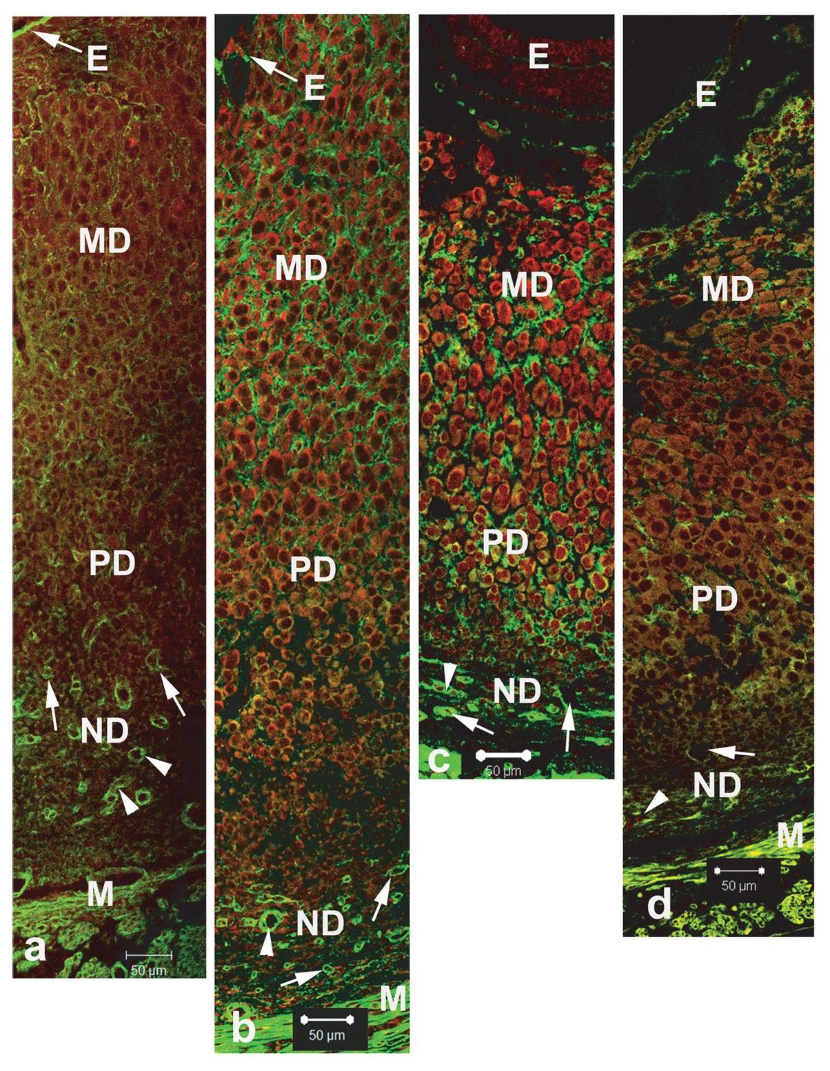 Fig 1. Spiess et al. Connective Tissue Research, 48:99–108, 2007.
Immunofluorescence for type III collagen in the antimesometrial decidua. (a) Day 5 of pregnancy. Immunofluorescence for collagen type III is present in mature decidua (MD) and predecidua (PD), forming a delicate network of fibers between decidual cells and in the nondecidualized region (ND). (b) Day 6 of pregnancy. Immunoreaction for collagen III is intense in the MD and PD forming a network of fluorescent fibers between decidual cells. In the PD the immunoreaction is less intense. (c) Day 7 of pregnancy: Collagen III is abundant through decidualized stroma and is observed as thick immunofluorescent fibers between decidual cells and predecidual cells. (d) Day 8 of pregnancy. Note that compared with day 7 the immunofluorescence is decreased in all decidualized stroma. In the ND, however, the immunofluorescence is maintained as in previous days and it is notable around blood vessels (arrow) and glands (arrow head). E = embryo, M = myometrium.
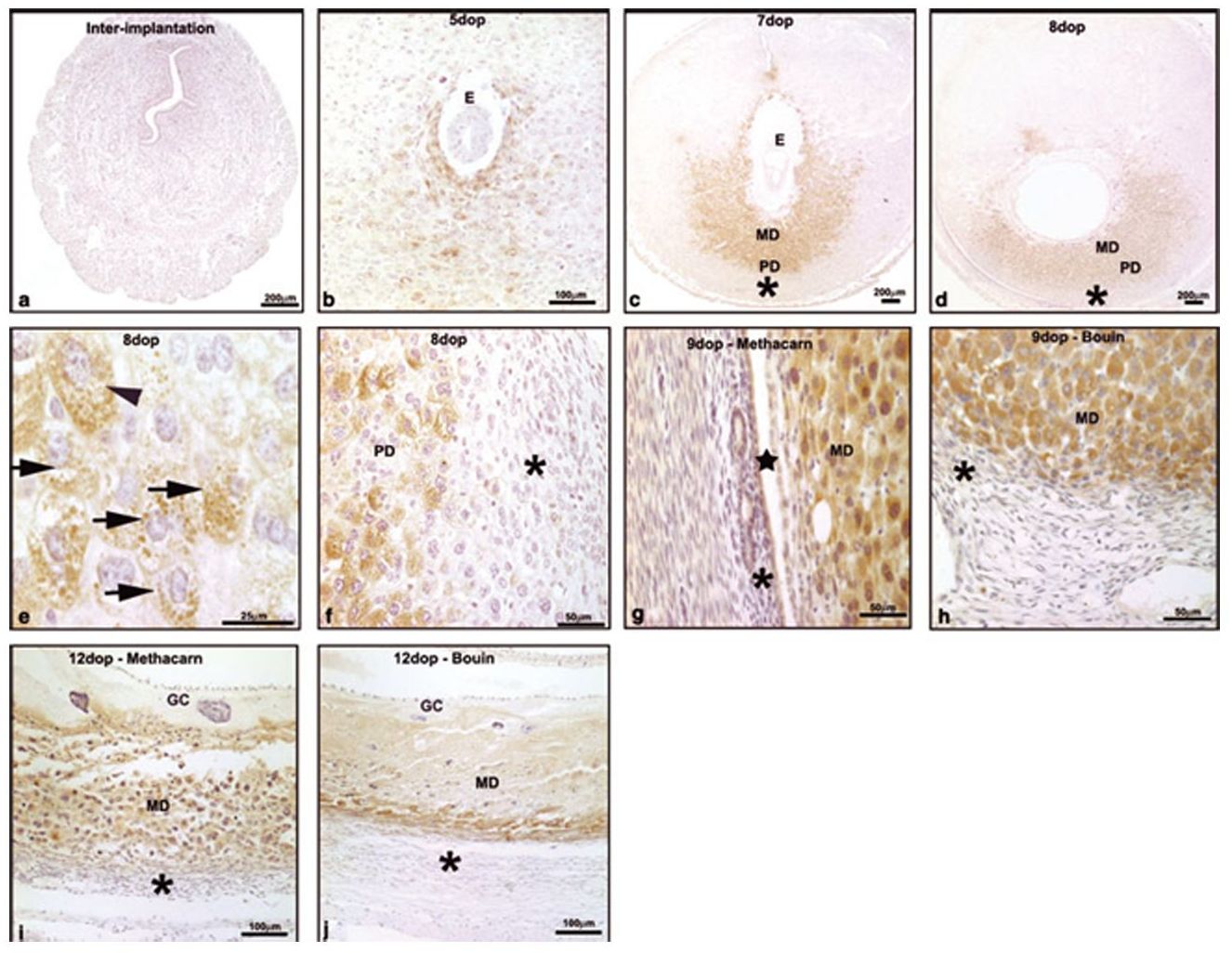 Fig 2. Cadeloro et al. Am J Reprod Immunol, 57:122–132, 2007.
Immunoperoxidase for decidual prolactin-related protein (dPRP) in the antimesometrial region: (a) no immunoreactivity at interimplantation sites; (b) day 5: dPRP is limited to the mature decidual cells surrounding the implantation crypt; (c, d) days 7 and 8: expanded decidualization, greater immunoreaction; (e) day 8: high magnification showing strong staining of cytoplasmic granules (arrows); (f) day 8: boundary between strongly labeled pre-decidual cells (PD) and non-labeled non-decidualized fibroblasts (asterisk); (g, h) day 9: dPRP-immunoreactive decidual cells (DC) in samples fixed with (g) methacarn or (h) Bouin’s solution; (i, j) day 12: immunoreactivity in the degenerating mature decidua (MD) in samplesfixed with (i) methacarn or with (j) Bouin’s solution. Star: new uterine lumen; asterisk: non-decidualized stroma.
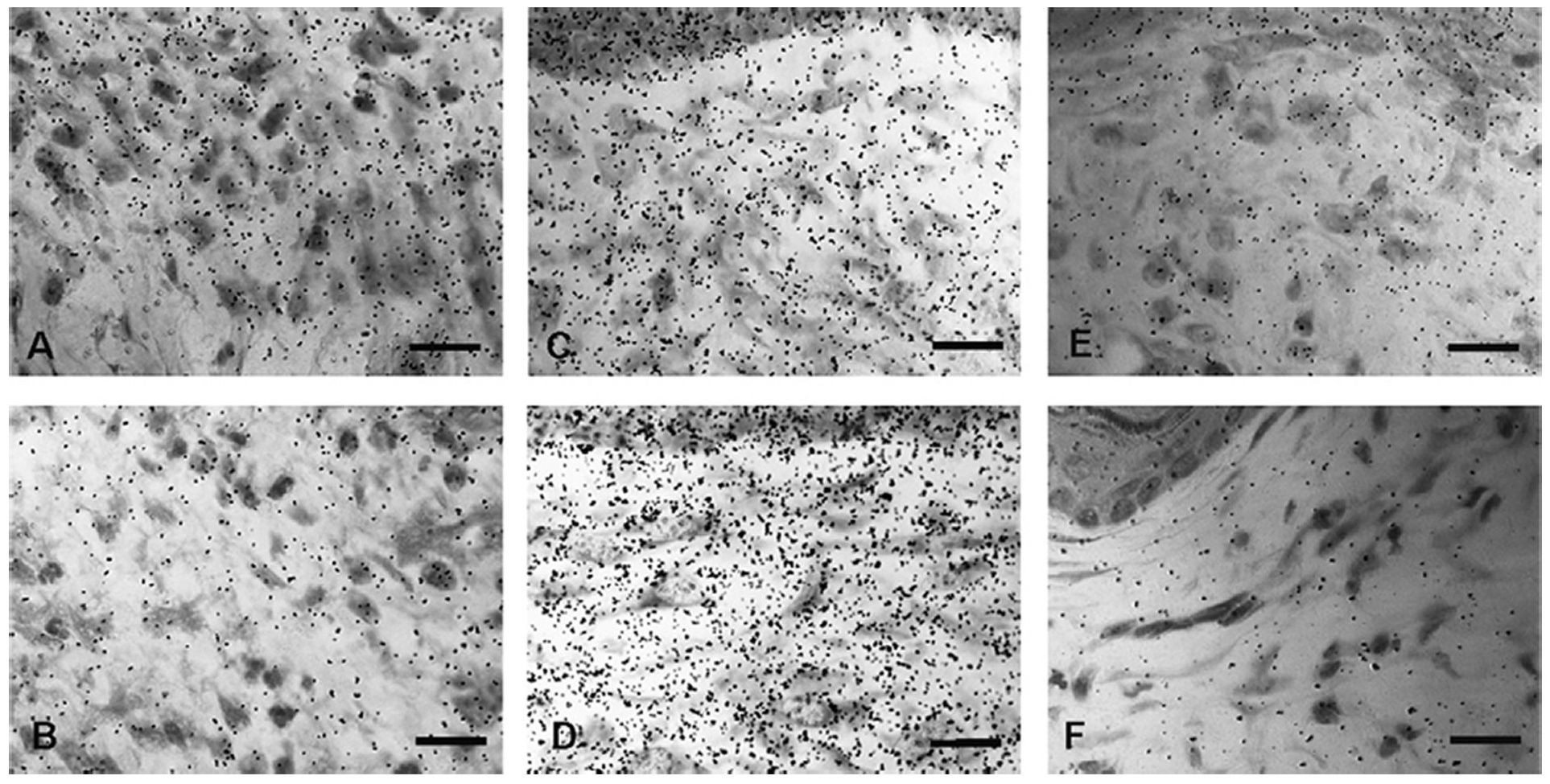 Fig. 3 Araujo et al. Journal of Photochemistry and Photobiology B: Biology 86: 87–96, 2007.
Autoradiograms after 3H-proline administration. (A) Control dermis on day 8 p.w. (B) Irradiated dermis on day 8 p.w. The fibroblasts are weakly labeled in both groups. (C) Control dermis on day 15 p.w. is higher labeled than day 8 p.w. (D) Irradiated dermis on day 15 p.w. show a higher 3H-proline incorporation than control lesions. Compare C and D. (E) Control dermis on day 22 p.w. (F) Irradiated dermis on day 22 p.w. The incorporation of 3Hproline decreases in both groups. Irradiated-lesions, however, show a lower incorporation than control lesions. Bar scale = 30 lm.
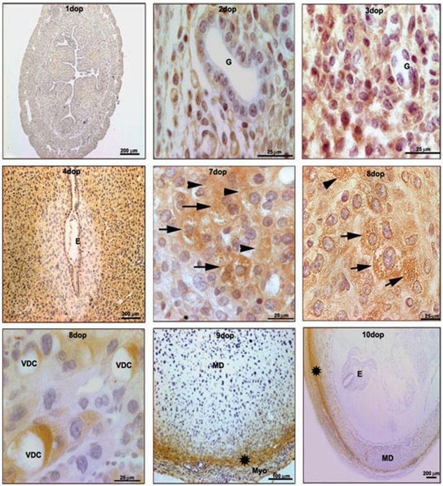 Fig. 4 Candelor et al. Am J Reprod Immunol 2007.
Immunoperoxidase for follistatin in the antimesometrial region: (a) no mmunoreactivityinterimplantation site; (b,c; days 2?3)labeled endometrial fibroblasts– unlabeledgland (G); (d; day 4) cells surrounding the implantation crypt unlabeled, whereas most peripheral cells are labeled; (e.g. days 7 and 8) labeled predecidual cells. The predecidualcells in these days showed different types of labeling. The immunoreaction was as a (e) diffuse cytoplasmic staining, (e and f), inside cytoplasmic granules (arrows) and (g) in the vacuoles periphery of the vacuolated decidual cells (VDCs). (h,i; days 9 ? 10) immunoreactivity restricted to non-decidualized cells (star).E: embryo; Myo: myometrium.
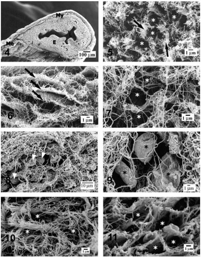 Fig 5 Carbone et al. Microscopy research and technique 69:36–45, 2006.
Scanning electron micrographs of mouse uteri digested with NaOH. Fig. 4: The main structural components of the uterus of a nonpregnant mouse can be recognized in a low magnification picture of a digested specimen: L, uterine lumen; E, endometrium; My, myometrium; Me, mesometrium. Fig. 5: Nonpregnant endometrium. The collagen fibrils form bundles or flat groups (arrows) that leave irregular spaces, formerly occupied by stromal cells (*). Fig. 6: Myometrium of a nonpregnant animal. The collagen fibrils form regular plates (arrows) that formerly surrounded the muscle cells. Fig. 7: Decidualized endometrium of a pregnant mouse. The collagen fibrils form delicate baskets whose spaces (*) were occupied by decidual cells.
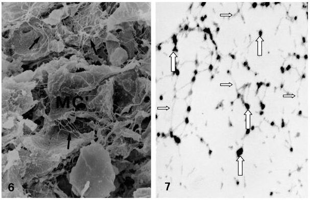 Fig 6 Pereda et al. Microscopy research and technique 69:386–395. 2006.
Image of a 5-weekold human embryo showing the migratory pathway of primordial germ cells. Note fine filamentous strands extend between the surfaces of the cells (arrows).MC, mesenchymal cells. 31,350. Fig. 7: Extracellular spaces of the migratory pathway of the primordial germ cells after treatment with ruthenium hexamine trichloride. Proteoglycans are observed by TEM as electron dense granules (large arrows) interconnected by thin filaments (small arrows). 324,600.
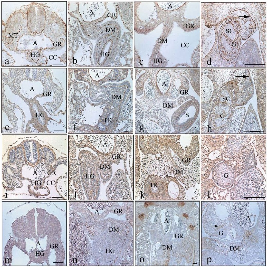 Fig. 7 SOTO-SUAZO et al. Histochem Cell Biol 121:149–153. 2004.
(a–p) Transverse hindgut (HG) sections of mouse embryos showing immunoperoxidase localization of the four glycoproteins: collagen type I (a–d), collagen type III (e–h), collagen type V (i–l), and tenascin-C (m–p). Arrows show basement membrane of the coelomic epithelium. DM Dorsal mesentery, GR genital ridge, G gonad, A aorta, CC coelomic cavity, MT mesonephric tubule, S stomach, SC sexual cord. Bars 100 mm
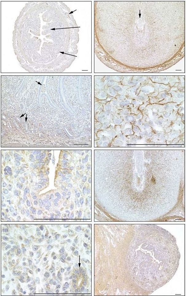 Fig. 8 San Martin et al. Reproduction 125, 585–595. 2003.
Immunoperoxidase localization of biglycan in the uterus in mice. (a) Day 1 of pregnancy. Biglycan is almost absent from all regions. (b) Longitudinal section of a uterine horn on day 4 of pregnancy. Biglycan is restricted to the deep stroma (DS) surrounding the glands and myometrium (My). (c) Day 5 of pregnancy. Immunoreaction for biglycan is observed in the cytoplasm of mature decidual (MD) cells and in the apical cytoplasm of epithelial cells that line the uterine crypt (*). (d) Day 5 of pregnancy. Note the reaction for biglycan in the cytoplasm of predecidual cells and uterine glands. (e) Day 6 of pregnancy. Reaction for biglycan is observed in the region of predecidual (PD) cells and deep stroma as well as in the myometrium, but not in mature decidua. (f ) High magnification of predecidual region showing biglycan-positive fibrils situated between the cells. (g) Longitudinal section of a uterine horn on day 7 of pregnancy. A
strong reaction for biglycan is now observed, particularly in the region of mature decidual cells and myometrium. (h) Longitudinal section of an interimplantation site (IIS) which is almost negative for biglycan. BV: blood vessel; E: embryo; G: uterine glands; L: uterine lumen;
M: mesometrium; SS: superficial endometrial stroma; T: trophoblast. Scale bars represent 100 ?m.
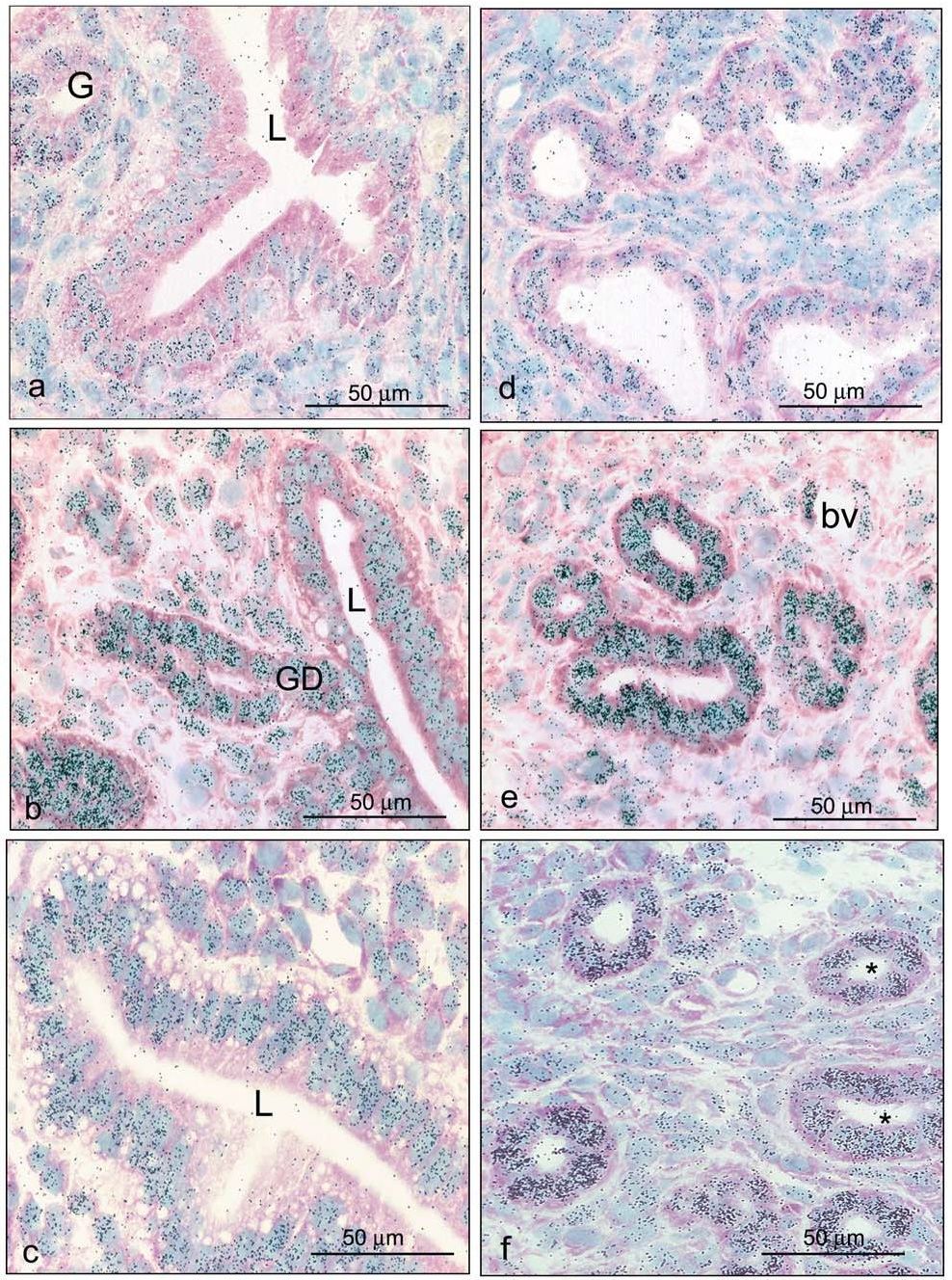 Fig. 9 zorn et al Histochem Cell Biol 120:1–12. 2003.
a–f 3H-estradiol concentration on days 1.5 (a, b), 2.5 (c, d),and 3.5 (e, f) after fertilization. In the luminal epithelium, nuclear labeling is higher at luminal branches and at mesometrial and antimesometrial extensions (a, c, e). The concentration of 3Hestradiol is higher in glands than in luminal epithelium (a–f).
Among glands, epithelial height, diameter of lumen, and nuclear silver grain density vary (c, d, f). Asterisks indicate low-labeled glandular profiles. G Gland, GD glandular duct, L uterine lumen, bv blood vessel. Exposure times: 70 days for c–f; 100 days for a, b.
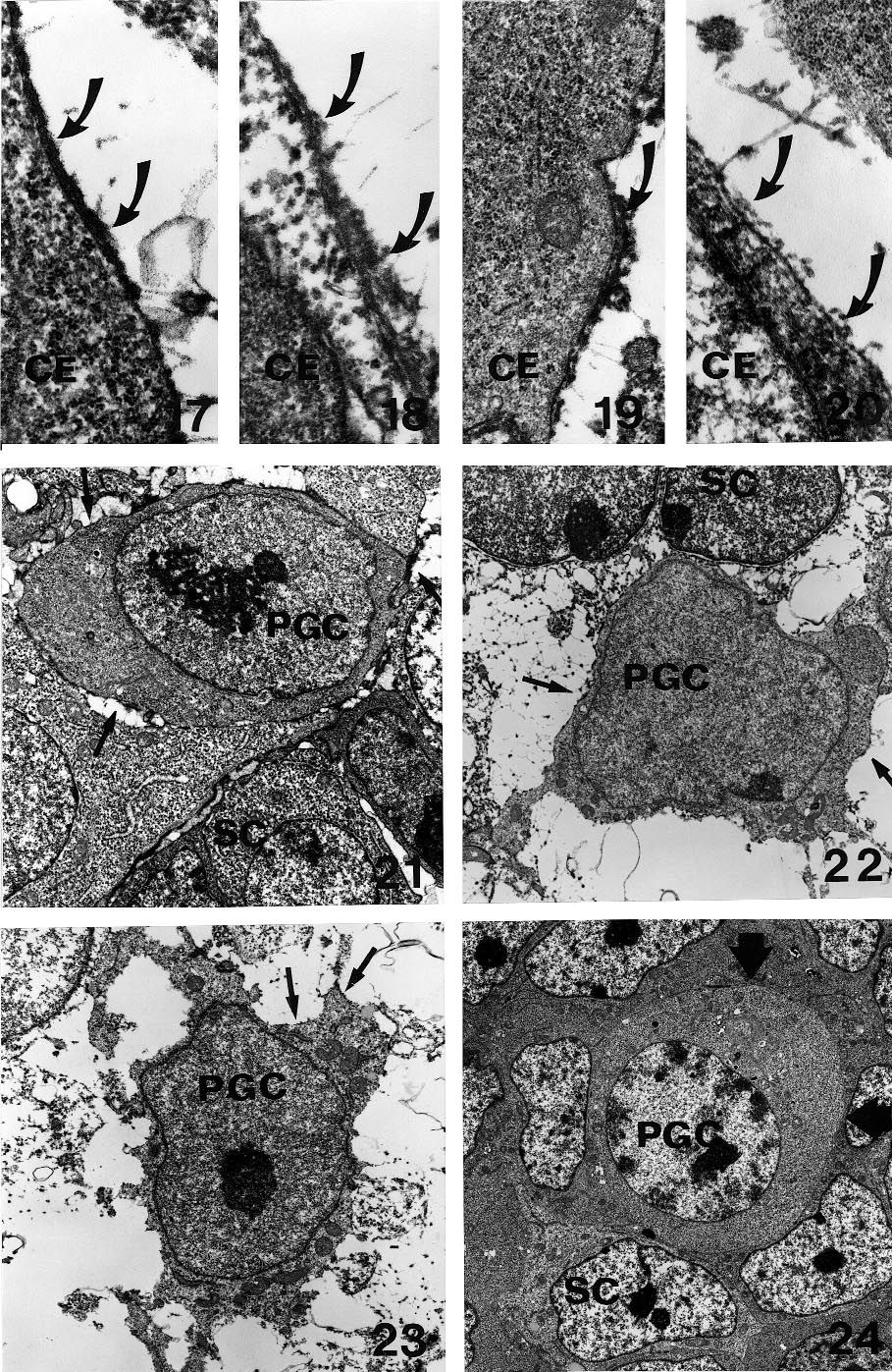 Fig. 10 Soto-Suazo et al. Tissue & Cell, 31 (3) 291–300. 1999.
Figs 17–20 Basement membranes of the coelomic epithelium (CE) of the migratory pathway. An electron dense RHT positive layer (arrows) is concentrated under the epithelial cells. This layer is more evident in 9- (Fig. 17) and 10-day embryos (Fig. 18) as compared with day 11-(Fig. 19) and day 12 embryos (Fig. 20). Electron micrographs. X 24 600. Figs 21–24 Primordial germ cells (PGC) treated with RHT. Observe a delicate RHT-positive reaction (arrows) on the surface of the of 9-, 10- and 11-day-old embryos (Figs 21–23). No reaction is observed on the surface of primordial germ cells (arrowheads) of 12-day-old embryos (Fig. 24). SC: Somatic cell. Electron micrographs.Figs 21–23: X 6150. Fig. 24: X 3100.