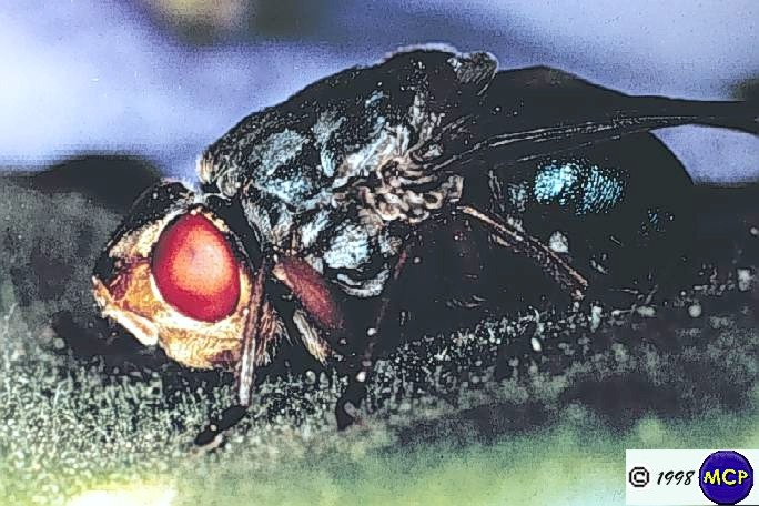
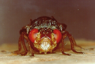
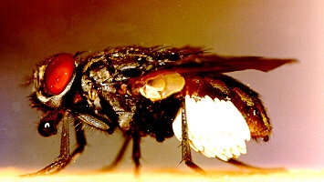
Dermatobia hominis is a bluebottle-like fly with yellow to orange head and legs. The thorax is dark blue with a greyish bloom; the abdomen is short and broad and has a brilliant blue color.
Adults have atrophied mouthparts and do not feed, relying instead on food reserves accumulated during the larval stage.
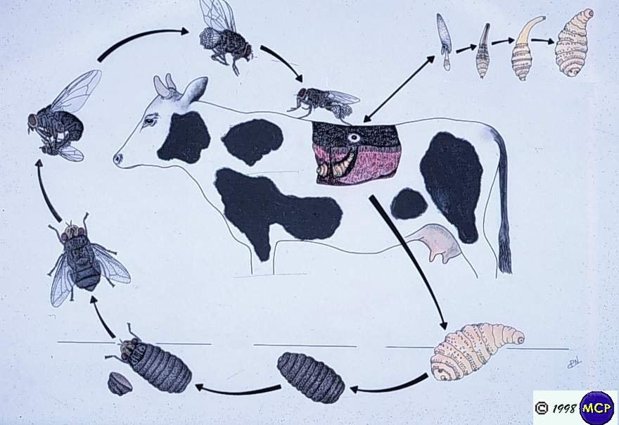
The length of the life cycle ranges between 100 and 120 days.
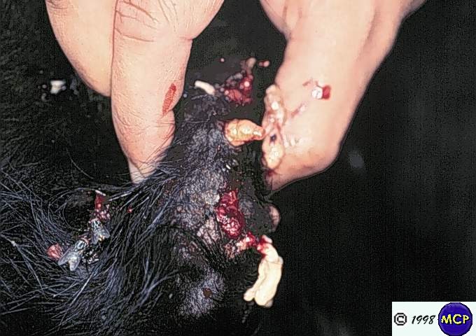
Each larva penetrates individually and a boil-like swelling develops around it - often at areas inaccessible to host grooming behavior. The swelling has an opening through which the larva respirates. Larval development is completed in 5-10 weeks, after which the larvae leave their host during the night or early morning, never during the afternoon - presumably to avoid risk of desiccation.
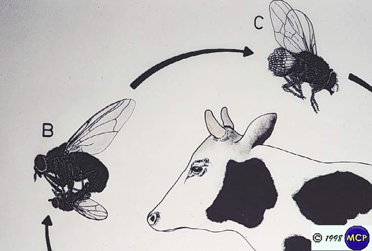
When the Dermatobia female is ready to oviposit she captures another insect - usually a fly - and glues her eggs to the captured insect's abdomen.
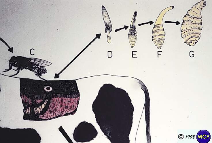
Development of the egg requires 4 to 9 days, but hatching is delayed until the stimulus of a sudden increase in temperature, which would occur when a carrier insect visits a warm-blooded host. The larva tranfers to the host, and either enters through the feeding puncture made by the carrier, or penetrates the unbroken skin, which can do on 5-10 minutes.
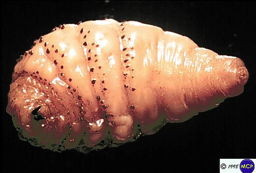
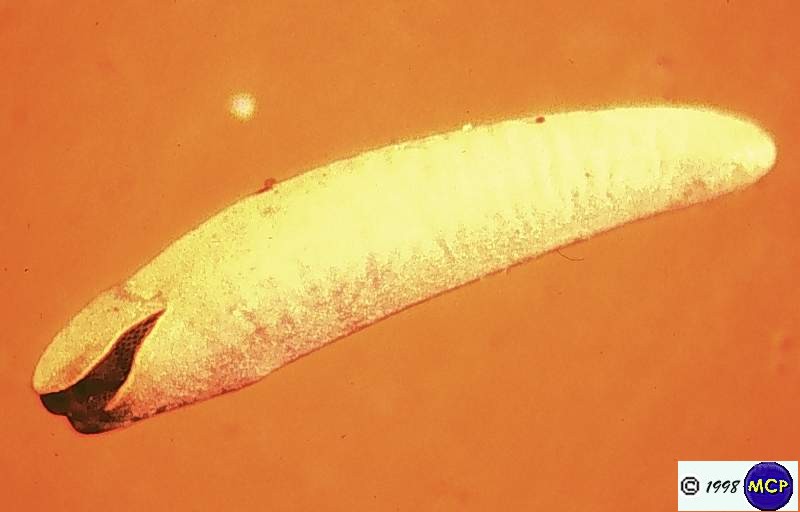
The third-stage larva is elongate ovate, with narrow belts of sparsely set spines, and prominent mouth-hooks. The posterior spiracles have three slits, no button and are sunk in a pit.
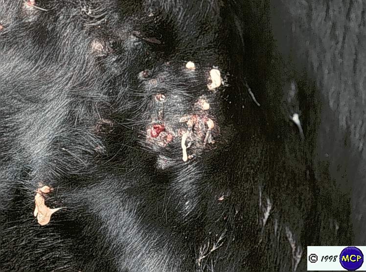
The larvae occur in swellings in various parts of the body, and these may suppurate and cause pain; in Brazil the condition is often known as berne. When the larva is removed, in the absence of secondary infection, the condition clears spontaneously in about a week.
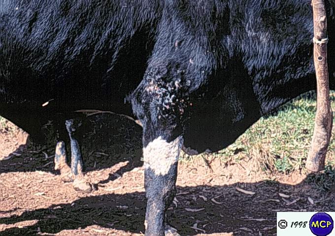
Dark colored areas of cattle are more attractive for blood-sucking phoretic day-flying species. Darker animals absorb more heat into the skin, and aldo radiate more; the higher skin temperature of dark-coloured cattle may be more attractive to blood-sucking insects .In Brazil it was found that 57% of Dermatobia nodules were located on the left side of the body. The hosts' preference for resting on their right side might be the reason for this asymmetric distribution of nodules: 55% rested on their right side so exposing their left side to carriers of Dermatobia eggs.
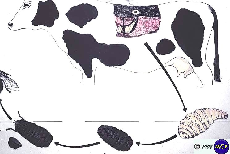
The mature larvae or prepupae emerge from the host after about three months and pupate on the ground for a further month before the adult fly emerge.
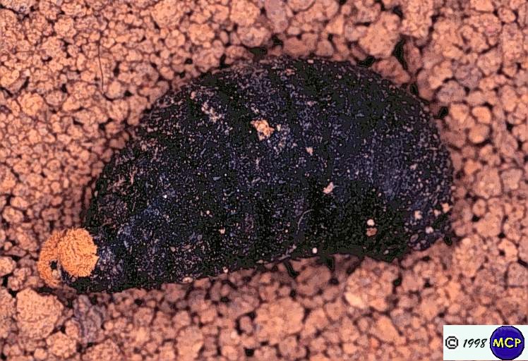
The puparium may exhibit prominent flower-like anterior spiracles of the third instar larva. The prepupae emerge from the host in the early morning, and burrow into the soil where they pupariate. The pupal stage is long, taking 4 to 11 weeks.
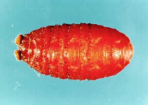
Most mature larvae drop during early morning hours. They burrow into upper soil or debris for about 20 min and form a hardened puparium in 2-3 days.