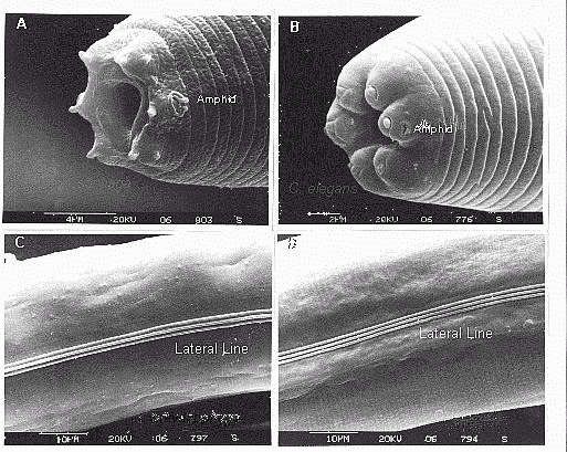
Scanning Electron Micrographs of Caenorhabditis elegans and
Oscheius tipulae
O. tipulae (A and C) and C. elegans (B and D) were
submitted to SEM to show their different external morphology. (A) and
(B) show the head and (C) and (D) show the lateral line. NOTE the
size and position of the amphids (chemosensory organs) and lips in both
species (A and B). NOTE also the different morphology of the lateral
line (C and D) in both animals.
These photos were kindly taken by R. Turner at the Department of
Biology, Indiana University, USA